The Hepatitis Delta neglected pandemic

Hepatitis is now a hot topic, due to the recent severe cases of liver inflammation in children between 1 month and 16 years of age in different countries. The first (and more numerous) cases have been reported in the United Kingdom, but also in Italy, Spain, Romania, United States, Israel, and other EU countries.

The cause of this acute paediatric hepatitis, that in some cases had lead to liver transplantation, is still unknown; moreover, it is not clear whether a real outbreak is taking place, or if the recent events had rase the awareness on this condition (when we actively look for something, we are more likely to find it). While some hypotheses are under study, including the involvement of adenovirus infection (all correlations with SARS-CoV-2 or with the COVI-19 vaccine have been discarded), the advice is to rely on respiratory and hand hygiene and to pay attention to symptoms such as fever, diarrhoea, abdominal pain, fatigue, dark urine, light-coloured stools, and jaundice (yellow discolouration of skin and eyes).

Hepatitis can be caused by viruses (as I mentioned here), by toxic substances (alcohol, excessive amounts of some drugs, fungal toxins, etc) or can be a symptom of autoimmune diseases (misdirected reactions of the immune system against some components of the human body).

Viral hepatitis is a very common disease, but often under-diagnosed and, as a consequence, not treated. This is particularly true in the case of hepatitis delta, caused by the combination of two viruses: hepatitis B virus (HBV) and hepatitis delta virus (HDV). In some cases both viruses infect a person at the same time (co-infection), while in others, a patient already infected by HBV can be later infected by HDV (super-infection). HBV-HDV double infection causes the most severe form of viral hepatitis that, if not treated, can lead to very severe consequences, including liver cancer. It is estimated that about 300 million people in the world are infected by HBV, and that 4,5% of them have been in contact with HDV, but real numbers might be even higher.

For both HBV and HDV transmission occurs through infected blood or other body fluids, or, especially in the areas where they are endemic, from mother to child at birth or early childhood.

Hepatitis delta virus, which I studied for 7 years, is a very interesting virus, with very peculiar characteristics:

  • It is the smallest virus known to infect humans
  • It has a RNA genome
  • Its replication occurs through mechanisms not seen in other human viruses
  • It shares similarities with some plant viruses (viroids)
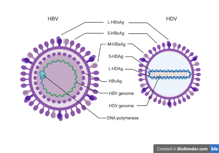
  • It has only two proteins (L-HDAg, S-HDAg), therefore depends almost entirely on human proteins
  • It can produce new viral particles and infect other cells, only in the presence of HBV
  • Its external layer (envelope) is made of HBV surface proteins.

This last characteristic has turned out to be very useful to limit the spread of the infection since the vaccine against HBV is highly protective also against HDV. If you, like me, were born in the 80s in Italy, you may remember receiving your HBV vaccination in the 90s, when it become compulsory. The HBV vaccination campaign was very successful and contributed to a strong reduction of the cases of hepatitis delta in Italy, where HDV was endemic and is now considered soon to be eradicated.

However, it is estimated that about 12 million people worldwide are currently infected with HDV, because in many areas vaccine coverage is still low; the number of people living with this virus can be even higher, since in many cases HDV diagnostic tests are not performed. (In the documentation about the recent cases of acute paediatric hepatitis, I have often read that the patients were negative for HBV, HCV, HAV, HEV, “and HDV if the test was available”).

Since HDV produces only two viral proteins, it relies on human proteins from the infected cells to replicate, therefore the development of specific antivirals has proven quite difficult (to block viral replication, we should block cellular proteins, with potentially severe side effects), and, unfortunately, the treatments commonly used against HBV are not effective against HDV.

New antivirals have recently been developed with different mechanisms of action to inhibit various steps of HDV infection, and some of them are giving promising results in clinical trials. In particular, two new drugs blocking respectively HBV and HDV viral entry in liver cells and blocking the formation of new HDV infectious particles have been conditionally approved in the United States and the European Union (approved before the end of clinical trials, because benefits are clearly largely overcome risks)

Despite HDV being discovered 45 years ago (in 1977 in Italy by Prof. Mario Rizzetto), there are still many things we do not know about how it causes the disease, and, most importantly, we still do not have a specific cure. Since HDV is more common in economically less developed countries, hepatitis delta have been for many years a neglected disease.

The conditional approval of the new drugs is a first step towards the improvement of the quality of life of patients with hepatitis delta, but more is needed: research must continue to fully understand what happens in the liver of infected patients, awareness about this disease needs to be raised to encourage prevention through vaccination and discourage high risk behaviours, and more resources are needed to ensure diagnosis in all suspected cases and improve vaccination coverage in developing countries.

Image created in BioRender.com by Carla Usai (L-HDAg, S-HDAg: HDV proteins; S-HBsAG, M-HBsAg, L-HBsAg: HBVsurface proteins; HBcAg: HBV core protein). Figure not drawn to scale

Bibliography:

Review article: emerging insights into the immunopathology, clinical and therapeutic aspects of hepatitis delta virus, Usai C et al., Alimentary Pharmacology and Therapeutics 2022 https://doi.org/10.1111/apt.16807

World Health Organisation page about acute severe hepatitis in children https://www.who.int/emergencies/disease-outbreak-news/item/multi-country-acute-severe-hepatitis-of-unknown-origin-in-children

I papillomavirus (HPV)

Quando parliamo di papillomavirus (HPV) non ci rifieriamo a un singolo virus, bensì a più di 200 virus diversi, tutti appartenenti alla famiglia dei Papillomaviridae, e classificati in 5 generi (α ,β, γ, μ, ν).

Si tratta di virus a DNA che infettano la pelle o le mucose, e vengono suddivis in tipi ad alto o a basso rischio in base alla loro capacità di causare il cancro. Si stima che 4 su 5 di noi siano entrati in contatto almeno una volta nella vita con un papillomavirus, spesso senza esserne consapevoli. Nella maggior parte dei casi, infatti, le infezioni da papillomavirus sono asintomatiche.

In generale, le infezioni da HPV non causano la morte delle cellule e il virus non circola mai nel sangue.

Nel caso dei papillomavirus a basso rischio l’infezione si manifesta con la formazione di verruche sulla pelle o papillomi nelle mucose: il virus infatti induce la proliferazione delle cellule epiteliali che spesso producono uno strato di cheratina che le riveste.

I papillomavirus ad alto rischio (ne sono stati individuati 15) possono causare lesioni più gravi, che se non curate adeguatamente possono trasformarsi in tumori causati dalla presenza continua del virus. I tessuti che possono essere colpiti da questi tumori si trovano nell vie aeree superiori e nei genitali sia maschili che femminili.

Harald zur Hausen fu il primo a dimostrare il ruolo degli HPV nella carcinogenesi, e per questo fu insignito del Premio Nobel per la Fisiologia e Medicina nel 2008.

Le lesioni benigne guarsicono spontaneamente, infatti in questi casi il sistema immunitario è capace di eliminare il virus senza che sia necessario alcun trattamento. A volte però le cellule che dovrebbero dare il via alla risposta immune falliscono nell’intento, e ciò fa sì che il sistema immunitario diventi tollerante al virus.

Gli HPV, a loro volta, si sono evoluti per sfuggire al sistema immunitario i diversi modi:

  • Non producono antigeni in grande quantità
  • Non uccidono le cellule infettate
  • Fanno diminuire la produzione di molecole infiammatorie
  • Limitano la presentazione delle proteine virali sulla superficie delle cellule infettate

Con questi stratagemmi gli HPV fanno in modo che il sistema immunitario non venga allertato della loro presenza e che l’infezioni possa diventare persistente. Ciò da tempo agli oncogeni (geni che causano tumori) del virus di modificare le cellule infettate e indurre la loro trasformazione in cellule tumorali, per esempio inducendo la fusione di cellule vicine, o bloccando l’ultima fase della divisione cellulare impedendo che due cellule figlie si separino. In poche parole, i virus prendono il controllo delle cellule infettate e ne alterano il metabolismo e il ciclo cellulare.

Tra tutti i papillomavirus conosciti, almeno 40 infettano i genitali, e papillomavirus ad alto rischio HPV-16 e HPV-18 causano da soli il 70% di tutti i casi di tumore alla cervice uterina.

La cervice uterina è la porzione inferiore dell’utero, che si divide a sua volte in due zone (esocervice ed endocervice) rivestite da due tipi di cellule diverse: le cellule squamose epiteliali e le cellule ghiandolari. A seconda del tipo di cellule colpito dall’infezione si possono sviluppare due tipi di tumori chiamati rispettivamente carcinoma a cellule squamose e adenocarcinoma.

L’infezione da HPV può essere diagnosticata con il Pap-test, un prelievo di cellule della cervice che permette di individuare eventuali cellule tumorali, o con l’HPV-test che determina l’eventuale presenza di DNA virale. La diagnosi precoce è fondamentale per eliminare l’infezione e bloccare lo sviluppo delle lesioni cancerose.

Negli ultimi anni, grazie alla diffusione di vaccini preventivi contro diversi tipi di HPV, il tumore alla cervice uterina è sceso dal secondo al quarto posto tra i più diffusi tra le donne, ma causa ancora il 6,5% di tutti i tumori diagnosticati nella popolazione femminile.

Gli HPV, in particolar modo HPV-16, possono causare anche carcinomi a cellule squamose dell’orofaringe secondo lo stesso meccanismo alla base del tumore alla cervice uterina: se il sistema immunitario non elimina il virus tempestivamente, l’infezione diventa persistente e il virus induce la trasformazione tumorale delle cellule. Tumori simili possono essere causati anche dall’esposizione a fumo e alcol indipendentemente da HPV (HPV-negativi), ma recentemente la proporzione di tumori dell’orofaringe HPV-positivi è aumentata notevolmente e si prevente che presto HPV ne diventerà la causa principale.

I carcinomi dell’orofaringe HPV-positivi sono più frequenti negli uomini che nelle donne.

Esistono tre vaccini preventivi contro i papillomavirus basati sulla proteina virale L1: molte unità di L1 si assemblano per formare delle particelle simili ai virus (VLP, virus-like particle) ma prive di DNA virale e di tutte le altre proteine, e pertanto non infettive e non oncogeniche. Il sistema immunitario riconosce le particelle simil-virali e inizia a produrre anticorpi contro L1, tra cui anche anticorpi neutralizzanti capaci di bloccare il virus e impedire l’infezione.

Il primo vaccino, autorizzato nel 2006, contiene le proteine L1 dei papillomavirus a basso rischio e ad alto rischio più comuni (HPV-6 e HPV-11; HPV-16 e HPV-18 rispettivamente). Nel 2009 venne approvato un secondo vaccini diretto esclusivamente contro HPV-16 e HPV-18. Il vaccino più recente venne approvato nel 2014 e comprende le proteine L1 di ben nove papillomavirus (HPV-6, HPV-11, HPV-16, HPV-18, HPV-31, HPV-33, HPV-45, HPV-52 e HPV-58).

Questi vaccini si sono dimostrati sicuri ed efficaci, prevenendo più del 90% delle infezioni, ma putroppo la loro diffusione nei paesi più poveri è ancora limitata, a causa degli alti costi di produzione e trasporto.

È importante ricordare che l’infezione da HPV colpisce entrambi i sessi, e che, per ridurre la diffusione dell’infezione a e l’incidenza dei tumori che essa causa (non solo il cancro alla cervice uterina), il vaccino è rivolto sia agli uomni che alle donne.

Immagine creata su BioRender.com da Carla Usai

Bibliografia

European Centre for disease prevention and Control https://www.ecdc.europa.eu/en/human-papillomavirus

An update on Human Papilloma Virus vaccines: history, types, protection and efficacy, Yousefi Z et al., Frontiers in Immunology 2022, https://doi.org/10.3389/fimmu.2021.805695

International standardization and classification of human papillomavirus types, Bzhalava et al, Virology 2022, https://doi.org/10.1016/j.virol.2014.12.028

Epidemiology of HPV-associated oropharyngeal cancer, Pytynia KB et al., Oral Oncology 2014, https://10.1016/j.oraloncology.2013.12.019

Global Cancer Statistics 2020: GLOBOCAN Estimates of Incidence and Mortality Worldwide for 36 Cancers in 185 Countries, Sung H et al., CA: A Cancer Journal for Clinicians 2021, https://doi.org/10.3322/caac.21660

Associazione Italiano Ricerca sul Cancro https://www.airc.it/cancro/informazioni-tumori/guida-ai-tumori/tumore-alla-cervice-uterina

Istituto Humanitas https://www.humanitas.it/malattie/infezione-da-hpv-papilloma-virus/

Human papillomavirus (HPV)

When we talk about human papillomavirus (HPV) we are actually referring to more than 200 different viruses, all belonging to the Papillomaviridae family, classified into five genera (α, β, γ, μ, ν).

They are DNA viruses that infect the skin or the mucosae and are divided into low-risk and high-risk types, depending on their ability to cause cancer. It is estimated that 4 out of 5 of us will be infected by an HPV at least once during our life, most of us without even realising it. In fact, in most cases papillomavirus infections are asymptomatic.

In general, HPV infections do not cause cell death, and the virus never spreads to the bloodstream.

In the case of low-risk papillomaviruses, the infection can manifest with the growth of warts on the skin or papillomas on the mucosae, because the virus induces the proliferation of epithelial cells that in turn produce a thick layer of keratine.

High-risk papillomaviruses (there exist at least 15 of them) can cause more severe lesions, that, if not treated, can evolve into tumours caused by the persistent presence of the virus. Such tumours can affect tissues in the upper respiratory tract and both male and female genitals.

Harald zur Hausen was the first to demonstrate the role of HPV in carcinogenesis, receiving the Nobel Prize for Physiology and Medicine in 2008.

Benign lesions caused by low-risk viruses heal spontaneously since the immune system is able to eliminate the virus without any medical treatment. However, sometimes, the cells responsible to start the immune response fail in their activity, and, as a result, the immune system grows tolerant to the virus.

In addition, HPVs have evolved different ways to evade the immune system:

  • They do not produce high quantities of viral antigen
  • do not kill the infected cells
  • inhibit the production of inflammatory signals
  • limit the presentation of viral proteins on the surface of the infected cells.

With these viral strategies in place, the immune system is not alerted of the presence of the virus, and the infection can go on unnoticed. This gives time to the oncogenes (tumour-inducing genes) to modify the infected cells and induce their cancerous transformation, for example inducing the fusion of neighbouring cells, or blocking the last phase of cell division preventing the two daughter cells to separate. Simply put, the virus takes the control of the infected cells and alters their cycle and metabolism.

At least 40 types of HPV infect genitals, and high-risk HPV-16 and HPV-18 alone cause 70% of all cervical cancer cases.

The uterine cervix is the lower portion of the uterus, and it can be divided into two parts (exocervix and endocervix) covered by two different cell types: epithelial squamous cells, and glandular cells. Depending on the cell type affected by the infection, two different types of tumour can develop, called squamous cell carcinoma and adenocarcinoma, respectively.

HPV infection of the cervix can be diagnosed through the Pap-test, the sampling of cells from the cervix to identify the presence of tumoural cells, or with the HPV-test, which determines the presence of viral DNA. Early diagnosis is essential to stop the development of cancerous lesions.

Thanks to the development and administration of prophylactic vaccines against some HPV types, cervical cancer is now the fourth most common cancer among women, after being the second one for a long time: however, it still causes 6,5% of all tumours diagnosed in the world female population.

HPV, particularly HPV-16, can also cause oropharyngeal squamous carcinoma, with the same mechanisms as cervical cancer: if the immune system fails to eliminate the virus early enough, the infection becomes persistent and the virus induces the tumoral transformation of cells. Similar tumours are also caused by exposure to smoke and alcohol independently of HPV (HPV-negative), but, recently, the proportion of HPV-positive oropharyngeal squamous cell carcinomas has increased and HPV is bound to become its main cause.

HPV-positive oropharyngeal squamous cell carcinomas are more frequent in men than women.

There are three approved vaccines against HPV, all based on the viral protein L1: many units of L1 assemble to form virus-like particles lacking viral DNA and all other viral proteins, and therefore not infectious and not oncogenic. The immune system recognises the virus-like particles and produced antibodies against L1, including neutralising antibodies able to block the virus and prevent infection.

The first vaccine, authorised in 2006, contains L1 proteins of the most common low-risk and high-risk HPVs (HPV-6 and HPV-11; HPV-16 and HPV-18, respectively). In 2009 a second vaccine was approved, exclusively directed against HPV-16 and HPV-18. More recently, in 2014, a third vaccine was approved, directed against 9 different types of HPV (HPV-6, HPV-11, HPV-16, HPV-18, HPV-31, HPV-33, HPV-45, HPV-52, and HPV-58).

These vaccines have proven safe and effective, preventing more than 90% of infections; unfortunately, their administration in low-income countries is still limited, due to the high production and transportation costs.

Do not forget that HPV infection affects both sexes, therefore, to reduce the viral spread and to decrease the incidence of HPV-caused tumours (not only cervical cancer), both men and women are eligible to receive the vaccine.

Image created in BioRender.com by Carla Usai

Bibliography

European Centre for disease prevention and Control https://www.ecdc.europa.eu/en/human-papillomavirus

An update on Human Papilloma Virus vaccines: history, types, protection and efficacy, Yousefi Z et al., Frontiers in Immunology 2022, https://doi.org/10.3389/fimmu.2021.805695

International standardization and classification of human papillomavirus types, Bzhalava et al, Virology 2022, https://doi.org/10.1016/j.virol.2014.12.028

Epidemiology of HPV-associated oropharyngeal cancer, Pytynia KB et al., Oral Oncology 2014, https://10.1016/j.oraloncology.2013.12.019

Global Cancer Statistics 2020: GLOBOCAN Estimates of Incidence and Mortality Worldwide for 36 Cancers in 185 Countries, Sung H et al., CA: A Cancer Journal for Clinicians 2021, https://doi.org/10.3322/caac.21660

Associazione Italiano Ricerca sul Cancro https://www.airc.it/cancro/informazioni-tumori/guida-ai-tumori/tumore-alla-cervice-uterina

Istituto Humanitas https://www.humanitas.it/malattie/infezione-da-hpv-papilloma-virus/

Mi presento

Ciao a tutti!

Mi chiamo Carla e sono una biotecnologa, da sempre affascinata dall’infinitamente piccolo e dal microscopico.

Per questo motivo mi interessano tanto i virus. Per diversi anni ho studiato il viurs più piccolo tra quelli che infettano gli esseri umani (il virus dell’epatite delta, HDV), e, più recentemente anche SARS-CoV-2, un virus di cui tutti abbiamo sentito parlare.

Attualmente lavoro a Barcellona, ma in passato ho lavorato anche a Madrid, Pamplona, San Diego e Londra.

Con questo blog mi propongo di mostrarvi il magico mondo dei virus e delle biotecnologie, e sarò felice di  farmi accompagnare da voi nelle mia vita quotidiana da ricercatrice. Troverete i miei post sia in Italiano che in Inglese.

Se avete delle curiosità o degli argomenti che vi interessano particolarmente lasciatemi un commento e sarete accontentati!

Photo credit: Carla Usai

Why don’t we all produce the same amount of antibodies against SARS-CoV-2?

What we already knew about the antibodies against SARS-COV-2

A negative antibody test (left) and a positive one (right). (“COVID-19-Test auf Antikörper IgG und IgM” by Partynia, from Wikimedia Commons under a A-S-A 4.0 International licence).

In our first study on patients who had recovered from COVID-19, we saw that some patients had more antibodies in their blood, and that in others the level of antibodies declined very quickly.

Moreover, looking at different groups of patients all over the world (including ours), it appears that about 10% of patients infected by SARS-COV-2 have detectable levels of antibodies against the N protein of the virus.

Our experiments

We decided to study the difference between those patients who recovered from COVID-19 and had detectable antibodies, and those who had not.

In particular, we analysed the blood of our patients looking at

  • the levels of neutralising antibodies, able to “capture” the virus and avoid the infection of more cells
  • the activity of immune cells called T cells in response to different proteins of the virus: Surface (S), Nucleocapsid (N), Membrane (M), and ORF-7a-2
  • the levels of molecules called cytokines and chemokines that, produced during infection, transmit messages between the infected cells and the immune system

Our results

  • We found that the majority of patients (about 70%) who did not have antibodies against the N protein, had neutralising antibodies that recognised the S protein. It is known that the production of antibodies against different viral proteins happens at different times during infection, and, like in waves, the levels of a new type of antibody start to increase when the previous ones start to fade. In our study, however, the patients with high levels of antibodies against N  antibodies had also high levels of neutralising antibodies against S, while those with low levels of anti-N had also low levels of neutralising antibodies.
  • We also found that T cells from different groups of patients have their own “favourite protein”: T cells from patients with antibodies against N react stronger against the S protein, while T cells from patients without antibodies against N, react against the Membrane protein (M), but not against S.
  • When we measured the levels of 20 cytokines and chemokines, we found that only one of them was different between the two groups: beta-NGF (β-NGF). Rita Levi Montalcini discovered this protein and its functions as a nerve growth factor in the 50s, and she was awarded the Nobel Prize for Physiology and Medicine in 1986.

What does β-NGF have to do with COVID-19?

This protein has been studied since the 50s, and it has been discovered that it serves several functions in the immune response, and that it is produced by B and T cells. Interestingly, its production increases during inflammation, and cells can respond to it using two different receptors:

  • TrkA induces cell activity and proliferation
  • p75 can cause cell death

When the levels of β-NGF in the blood are high, some cells produce more TrkA to be able to respond more efficiently to it. The interaction between β-NGF and TrkA has already been studied in the context of other respiratory diseases caused by viruses, like the respiratory syncytial virus (RSV) and human rhinovirus (HRV).

We found that those patients who did not produce antibodies against the N protein had lower levels of β-NGF, and their T cells had lower amounts of TrkA, and were, therefore, less active. This difference was particularly relevant in one subtype of T cells that help B cells produce antibodies. It seems, therefore that the message transmitted by β-NGF and received by TrkA is very important also for the immune response against SARS-CoV-2.

Why are these results important?

This is only one of the possible differences between people producing different amounts of antibodies against SARS-CoV-2, and the fact that the immune response against this virus does not have the same intensity or the same duration in all of us is surely the result of many factors. We need more studies with more patients of different ethnicities to complete this huge puzzle.

Understanding why the immune response is so variable in all its components – not only antibodies –  is extremely important to organise the vaccination programs and decide which groups are more at risk and should be considered the top priority. Vaccines are still our most valuable tool to prevent severe COVID-19 and to limit the spread of the virus, and they should be used in the wisest possible way.

Bibliography

The β-NGF/TrkA signalling pathway is associated with the production of anti-nucleoprotein IgG in convalescent COVID-19, Usai C et al., Frontiers in Immunology 2022 https://doi.org/10.3389/fimmu.2021.813300

Perché alcune persone non producono anticorpi contro SARS-CoV-2?

Nel nostro primo studio su pazienti guariti da COVID-19, abbiamo visto che alcuni pazienti producevano più anticorpi di altri, e che in alcune categorie questi anticorpi diminuivano più rapidamente.

Un test sierologico negativo (a sinistra) e uno poistivo (a destra). (“COVID-19-Test auf Antikörper IgG und IgM” di Partynia, da Wikimedia Commons con licenza A-S-A 4.0 International).

Inoltre, analizzando diversi gruppi di individui convalescenti in molte parti del mondo (compreso il nostro), è stato notato che circa il 10% delle persone che avevano avuto l’infezione avevano livelli non rilevabili di anticorpi contro la proteina N del virus.

Ci siamo posti l’obiettivo di capire che differenza ci fosse tra i pazienti guariti da COVID-19 che producevano anticorpi e quelli che invece non ne producevano.

Per fare ciò abbiamo analizzato nel sangue dei nostri pazienti

  • i livelli di un altro tipo di anticorpi, ovvero gli anticorpi neutralizzanti (quelli capaci di “catturare” il virus e impedire l’infezione di altre cellule).
  • l’attività dei linfociti T in risposta a “pezzettini” di diverse proteine del virus (S, N, M e una proteina non meglio caratterizzata chiamata ORF-7a-2).
  • i livelli di alcune sostanze prodotte in caso di infezione e infiammazione chiamate citochine e chemochine, che fungono da messaggeri tra le cellule infettate e le cellule del sistema immunitario.

Per prima cosa abbiamo trovato che 5 su 7 pazienti che non avevano anticorpi anti-N avevano però anticorpi neutralizzanti (che riconoscono soprattutto, ma non unicamente, pezzettini della proteina S). Questo non ci deve sorprendere, perché è già noto che la produzione di anticorpi contro proteine diverse del virus avviene con tempistiche diverse e con diversi andamenti nel tempo. In genere però, gli individui con alti livelli di anticorpi anti-N avevano anche alti livelli di anticorpi neutralizzanti, mentre quelli con bassi livelli di anti-N avevano bassi livelli di anticorpi neutralizzanti.

L’attività dei linfociti T, invece, sembra essere indipendente dalla presenza o meno di anticorpi. Infatti, molti studi hanno trovato che anche in assenza di anticorpi, i linfociti T di pazienti guariti dal COVID-19 sono in grado di reagire contro pezzi di proteine del virus, e l’intensità di questa risposta non dipende dal livello di anticorpi presenti nel sangue. Anche noi abbiamo ottenuto risultati simili, ma il risultato più interessante è che i linfociti T dei due gruppi di pazienti sembravano avere ognuno la sua proteina “preferita”: mentre i linfociti T degli individui che producevano anticorpi contro N reagivano più intensamente e meglio contro porzioni della proteina S, quelli che non li producevano reagivano molto bene contro porzioni della proteina di membrana (M), ma non reagivano alla proteina S.

Quando abbiamo misurato i livelli di 20 citochine e chemochine nel sangue, abbiamo visto che solo una era diversa tra i due gruppi: beta-NGF (β-NGF). La scoperta di questa proteina negli anni ‘50 e della sua funzione necessaria per far crescere le cellule nervose, valse il Premio Nobel per la Fisiologia e la Medicina a Rita Levi Montalcini nel 1986. Ma cosa c’entra con il COVID-19?

Dagli anni ’50 in poi questa proteina è stata molto studiata, e si è scoperto che ha molteplici funzioni anche nell’ambito del sistema immunitario, e che può essere prodotta anche dai linfociti B e T. In particolar modo la produzione di questa sostanza aumenta durante l’infiammazione, e può essere riconosciuta dalle cellule attraverso due recettori diversi, uno che fa aumentare l’attività e la proliferazione delle cellule (TrkA) e uno che invece può causare la morte cellulare (p75). Inoltre, quando i livelli di β-NGF nel sangue sono elevati, alcune cellule producono più TrkA per essere capaci di rispondere al meglio a questo segnale.

La comunicazione tra cellule che avviene attraverso β-NGF e il suo recettore TrkA è già stata studiata nel caso di malattie causate virus che infettano le vie respiratorie, come il virus respiratorio sinciziale (RSV) e il rinovirus (HRV).

Noi abbiamo trovato che i soggetti che non producevano anticorpi anti-N avevano livelli più bassi di β-NGF nel sangue, e per di più i loro linfociti T avevano meno molecole di TrkA. Questa differenza risultava particolarmente significativa per una categoria di linfociti T, i cosiddetti CD4+, che hanno l’importante compito di aiutare i linfociti B a produrre anticorpi. Sembra quindi che il messaggio trasmesso da β-NGF e ricevuto da TrkA sia molto importante anche nel caso della risposta immunitaria contro SARS-CoV-2.

Questa è solo una delle tante differenze tra le persone che producono più o meno anticorpi contro SARS-CoV-2, e ci sono sicuramente tanti altri fattori che fanno sì che la risposta immunitaria contro SARS-CoV-2 non abbia la stessa intensità o durata per tutte le persone che sono state infettate. Sono necessari altri studi con gruppi di pazienti più numerosi e di diverse etnie per chiarire questo fenomeno e aggiungere altre tessere a questo enorme puzzle.

Capire a cosa sia dovuta la variabilità della risposta immunitaria in tutte le sue componenti (ricordiamoci che non esistono solo gli anticorpi!) è fondamentale per gestire al meglio i programmi di vaccinazione stabilendo a quali gruppi dare la priorità. I vaccini restano la nostra arma migliore per prevenire forme gravi di COVID-19 e per limitare la diffusione del virus.

Bibliografia

The β-NGF/TrkA signalling pathway is associated with the production of anti-nucleoprotein IgG in convalescent COVID-19, Usai C et al., Frontiers in Immunology 2022 https://doi.org/10.3389/fimmu.2021.813300

Il fascino dell’Immunologia

La settimana scorsa ho partecipato al congresso della Società Britannica di Immunologia nell’incantevole Edimburgo.

Finalmente un vero congresso in presenza (era disponibile anche la modalità online per chi lo preferisse) con la possibilità di interagire con colleghi provenienti da tutto il Regno Unito e anche qualche ospite internazionale.

Quando partecipo a congressi così grandi che prevedono sessioni parallele su argomenti diversi, cerco sempre di seguire qualche sessione che non sia direttamente collegata al mio lavoro, per imparare qualcosa di nuovo.

Questa volta sono rimasta molto colpita dalla sessione sull’orologio biologico del sistema immunitario. A quanto pare le cellule del sistema immunitario preferiscono migrare dal sangue verso i linfonodi durante la notte, per presentare ai linfociti gli antigeni che hanno incontrato durante il giorno. Ciò significa che c’è un momento della giornata migliore per essere vaccinati o per ricevere una terapia che agisce sul sistema immunitario.

Anche il momento della giornata in cui un organismo si nutre, e dunque attiva certi meccanismi del suo metabolismo hanno un effetto sull’attività del sistema immunitario, per non parlare del fatto che il tipo di dieta che assumiamo influenza la composizione dei batteri che vivono nel nostro intestino (flora intestinale o microbiota intestinale), che a sua volta può modificare i livelli di molecole pro e anti infiammatorie nel sangue e nei tessuti.

Un altro fatto molto interessante è che infezioni passate o in corso possono modificare il risultato di un’infezione da parte di un microorganismo completamente diverso. Per esempio, sapevate che la presenza di un parassita nell’intestino (Heligmosomoides polygyrus) può favorire la produzione di un certo tipo di globuli bianchi nel midollo osseo, e quindi aiutare la risposta immunitaria contro il virus respiratorio sinciziale (RSV) che infetta i polmoni? O che la malaria (causata dal parassita Plasmodium falciparum che vive nei globuli rossi o nel fegato a seconda della fase di sviluppo) può causare un disequilibrio della flora intestinale, rendendo i bambini più soggetti a infezioni da batteri del genere Salmonella?

Inoltre, si stima che durante la pandemia di Influenza spagnola (di cui ho parlato qui), la maggior parte delle morti sia stata causata da un batterio (Streptococco pneumoniae) che approfittava dell’organismo già debilitato dall’infezione virale per danneggiare ulteriormente i polmoni, piuttosto che dal virus in sé. E recentemente è stato scoperto che una precedente infezione batterica da Klebsiella pneumoniae limita l’attivazione delle cellule del sistema immunitario chiamate Natural Killer, rendendo meno pericolosa una successiva infezione da parte del virus dell’influenza perché proprio queste cellule sono in parte responsabili del danno ai polmoni dovuto a un’eccessiva risposta immunitaria.

Insomma, ancora una volta sono rimasta affascinata dalla complessità del sistema immunitario e dalla molteplicità dei meccanismi con cui interagisce con praticamente ogni altro processo che avviene nel nostro organismo!

Nell’ultima sessione del congresso, invece, il Professor Paul Moss dell’Università di Birmingham e la Professoressa Sarah Gilbert dell’Università di Oxford, hanno ripercorso tutte le scoperte e i progressi fatti dalla scienza negli ultimi due anni: dal primo caso di polmonite atipica in Cina fino al richiamo del vaccino per la prevenzione della COVID-19. Probabilmente nessuno tra i presenti al congresso, neanche il più esperto tra gli immunologi, sarebbe stato così ottimista da prevedere che si sarebbero ottenuti così tanti risultati in così poco tempo.

Ed è proprio grazie a questo lavoro incredibile e alla collaborazione tra scienziati da ogni angolo del mondo che abbiamo avuto la possibilità di godere di questi fantastici quattro giorni all’insegna dell’Immunologia, e di visitare una città incantevole come Edimburgo (ovviamente muniti di COVID-pass e mascherina!)

Immagine da cellcartoons.net con licenza Creative Commons BY-NC-ND.

Immunology is cool!

Last week I attended the British Society for Immunology Congress in lovely Edinburgh.

At last a “real” in-person congress (the online option was also available) with real interactions with other scientists from all over the United Kingdom and some international guests.

When I take part in big congresses like this, with parallel sessions on different topics, I make time to attend some of the sessions not strictly related to my research, to learn something new.

This time, I have particularly enjoyed the session about the biological clock of the immune system. It looks like that the immune cells migrate from the blood to the lymph nodes during the evening, to present the antigens they have encountered during the day to other immune cells. This means that there is an ideal time of the day to get a vaccine or any other therapy that affects the immune system.

Also, the time at which an organism eats, and therefore activates certain mechanisms in its metabolism, has an effect on the activity of the immune system, and the type of diet can alter the composition of the bacteria that live in our gut (intestinal flora or microbiota), which in turn can modify the levels of pro- and anti-inflammatory signals in our blood and tissues.

Another fascinating fact is that current or previous infections can modify the outcome of an infection by a completely different microorganism. For example, did you know that the presence of a parasite in the intestine (Heligmosomoides polygyrus) can promote the production of a certain type of white cells in the bone marrow, and help the immune response against the respiratory syncytial virus (RSV) that infects the lungs? Or that malaria (caused by the parasite Plasmodium falciparum living in red blood cells or liver cells depending on the phase of its development) can cause an imbalance in the intestinal flora, increasing the risk for children of being infected by Salmonella bacteria?

What’s more, it seems that during the Spanish flu pandemic (mentioned here), most of the deaths were caused by a bacterium (Streptococcus pneumoniae) that took advantage of the organisms already weakened by the viral infection to further damage the lungs, and not to the virus itself. And recently, it has been discovered that a previous infection by bacterium Klebsiella pneumoniae can limit the activation of the immune cells called Natural Killer cells, making a later influenza virus infection less dangerous, since Natural Killer cells are partly responsible for lung damage in an excessive immune response.

In other words, I am more than ever in awe of the complexity of the immune system and of the many mechanisms through which it interacts with pretty much all processes happening in our body!

In addition, in the last part of the congress, Professor Paul Moss from the University of Birmingham and Professor Dame Sarah Gilbert from the University of Oxford went through all the discoveries and progresses made by the scientific community during the last two years: from the first reported cases of atypical pneumonia to the administration of the COVID-19 booster jab. I think that nobody, including the most expert immunologist, was so optimistic at the beginning of the pandemic to anticipate so many important results in such a short time!

And it was thanks to the amazing effort and collaborations between scientists all over the world, that we were able to enjoy those four intense days about Immunology and to visit the charming city of Edinburgh (with a COVID pass and the mask on, of course!)

Image from cellcartoons.net under Creative Commons BY-NC-ND licence.

Florence Nightingale: una pioniera dell’epidemiologia e del controllo delle malattie infettive

Florence Nightingale era un’infermiera dell’Inghilterra vittoriana, anzi sarebbe meglio dire che fu la prima vera infermiera. Fu lei infatti a definire l’infermieristica come professione (all’epoca e per molti anni riservata unicamente alle donne) e a codificarne principi e regole nel suo saggio Notes on nursing for the labouring classes (1868).

Nata in una famiglia dell’alta borghesia, come molte ragazze della sua classe sociale, si dedicava ad attività caritatevoli, tra cui l’assistenza ai malati. Inizialmente lavorò come volontaria in un piccolo ospedale londinese accumulando esperienza nella gestione e assistenza ai malati, ma la svolta avvenne durante la Guerra di Crimea (1853-1856) quando fu autorizzata a recarsi all’ospedale militare di Scutari, in Turchia, a capo di un gruppo di infermiere.

Dotata di un forte spirito di osservazione e attenzione ai dettagli, si rese conto che nella maggior parte dei casi i soldati morivano non per le conseguenze delle ferite di guerra, ma a causa di malattie contratte nell’ospedale stesso. Mise in atto un sistema di osservazione e raccolta dettagliata di informazioni quotidiane per ogni soldato che si trovasse nell’ospedale, tenendo traccia di sintomi, reazioni alle cure e modalità e tempi dei decessi. In Notes on Nursing scrisse che la caratteristica più importante dell’infermiera è l’abilità di osservare e riportare cambiamenti nelle condizioni dei pazienti. Fu quindi la prima ad applicare il metodo scientifico e la statistica in questo contesto.

Elaborò un nuovo tipo di grafico chiamato a rosa o coxcomb, l’antenato del grafico a torta che tutti conosciamo, in cui rappresentò “le cause di morte nell’esercito in Oriente”.

All’epoca la teoria dei germi (secondo cui le malattie vengono trasmesse dai microbi) non era ancora diffusa, e si riteneva che le malattie fossero trasmesse attraverso aria insalubre contenente particelle velenose (teoria dei miasmi). Nightingale si rese conto che sovraffollamento, scarsa ventilazione e mancanza di igiene erano la cause principale dei decessi che avvenivano nell’ospedale. Riorganizzò la disposizione dei letti, e diede alle sue infermiere istruzioni precise su come e con quale frequenza dovessero lavarsi le mani; si impegnò affinché i bendaggi e le coperte venissero lavati tra un paziente e l’altro.

Secondo Nightingale, le condizioni ambientali erano fondamentali nel determinare lo stato di salute dei pazienti, identificando come fattori principali la presenza di acqua pulita, aria pura, luce, igiene e un sistema fognario efficiente. Le innovazioni da lei introdotte contribuirono a far diminuire del 20% la mortalità tra i soldati.

Basandosi sull’esperienza nella Guerra di Crimea e in altri ospedali, scrisse il saggio Notes on Hospitals (1863) in cui descriveva la struttura ideale degli ospedali, ovvero con padiglioni che si diramano da un corridoio centrale per massimizzare luminosità e ventilazione, e minimizzare il contatto tra pazienti affetti da malattie diverse. Questa struttura fu per anni il modello sui cui si basò la costruzione di molti ospedali.

La sua passione e capacità per la matematica e la statistica la portò ad essere la prima donna ammessa nella Royal Statistiscal Society nel 1858.

Il contributo di Florence Nightingale alla statistica, epidemiologia, e controllo delle malattie infettive è stato talmente importante che l’Organizzazione Mondiale della Sanità decise di proclamare il 2020, bicentenario della sua nascita, Anno Internazionale dell’Infermiere. In quel momento non si sarebbe potuto prevedere che proprio nel 2020 gli infermieri e tutti i professionisti della sanità avrebbero avuto un ruolo fondamentale, e che i principi descritti per la prima volta da Florence Nightingale sarebbero stati di primaria importanza nella gestione della pandemia. Per celebrare ulteriormente questa eccezionale figura e la rilevanza del suo lavoro, il sistema sanitario nazionale britannico (NHS) decise di intitolare a Florence Nightingale gli ospedali di emergenza allestiti durante i primi mesi della pandemia per fronteggiare l’elevato numero di ricoveri.

Quasi 170 anni dopo la pubblicazione degli scritti di Nightingale, che ignorava cosa fosse un virus, ci siamo ritrovati ad applicare quotidianamente i concetti da lei postulati: lavarsi le mani, evitare sovraffollamenti, ventilare i locali.

Campagna dell’NHS per prevenire la diffusione del SARS-CoV-2 (2021)

Immagine principale: Diagramma di Florence Nightingale, Dominio Pubblico, tramite Wikimedia Commons

Bibliografia

Notes on Nursing for the Labouring Classes, Florence Nightingale; Harrison 18684

Notes on Hospitals, Florence Nightingale; Longman, Roberts, and Green 1863

Florence Nightingale’s Environmental Theory and its influence on contemporary infection control, Gilbert HA, Collegian 2020 https://10.1016/j.collegn.2020.09.006

Florence Nightingale’s lasting legacy for health care, Tye J, Nurse Leader 2020 https://doi.org/10.1016/j.mnl.2020.03.023

The art of medicine. Celebrating Florence Nightingale’s bicentenary, McEnroe N, The Lancet 2020 https://doi.org/10.1016/S0140-6736(20)30992-2

Florence Nightingale: a pioneer in epidemiology and infectious disease control

Florence Nightingale was a nurse during the Victorian age in England, or to be more precise, she was the first real nurse. She was the first to define nursery as a profession (for a long time exclusively for women), and to define its rules and principles in her book Notes on nursing for the labouring classes (1868).

A member of the upper class, she was involved in charitable activities, and he worked as a volunteer in a small hospital in London, where she learnt how to manage and assist patients. The turning point in Florence Nightingale’s life was the Crimean War (1853-1856), when she went to the military hospital in Scutari, Turkey, at the head of a group of nurses.

Thanks to her strong attention to detail, she observed that in most cases British soldiers were not dying of wounds acquired on the battlefield, but of illness contracted in the hospital. She set up a system of daily observation and detailed record of data from every patient, tracking symptoms, reaction to cure, time and modality of death. In Notes on Nursing, she wrote that the most important characteristic of a nurse should be the ability to observe and record any changes in patient conditions. She was the first to apply statistics and scientific method to this context.

She invented a new graph called the rose chart or coxcomb, ancestor of the pie chart we all know, in which she represented “the causes of mortality of the Army in the East”.

The germ theory (stating that diseases are spread by microbes) was not widespread at that time, and it was thought that diseases spread through venomous particles in the air (the miasm theory). Nightingale realized that overcrowding, scars ventilation and scars hygiene were the major causes of death in the hospital. She reorganized the distribution of the beds, and gave precise instructions on how and how often nurses needed to wash their hands; she insisted that bandages and blankets had to be washed between patients.

In Nightingale’s theory, the environmental conditions were of primary importance to determine the health status of the patients and identified clean water, pure air, light, cleanness and an efficient drainage system as major factors. Her innovations contributed to a 20% decrease in mortality among soldiers.

Based on her experience during the Crimean War and in other hospitals, she wrote Notes on Hospitals (1863), where she described the ideal architecture of hospitals: pavilions branching out from a central hallway to maximize light and ventilation, and to minimize contact between patients with different diseases. For many years new hospitals were built according to her model.

Gifted with passion and ability in mathematics and statistics, she was the first woman to be admitted to the Royal Statistical Society in 1858.

Florence Nightingale’s contributions to statistics, epidemiology and infectious disease control were so important that the World Health Organisation declared 2020, the bicentenary of her birth, the International Year of the Nurse. At that moment, it could have been impossible to imagine that in 2020 nurses and all the healthcare workers were bound to have such a critical role and that the principles introduced for the first time by da Florence Nightingale would have been of extreme importance in the fight against the pandemic. To further celebrate this extraordinary figure, the National Health Service decided to name after Florence Nightingale the temporary hospitals built as an emergency response during the first months of the pandemic to face the huge number of hospital admissions.

Almost 170 years after the publication of Nightingale’s books, when the concept of a virus was pretty much unknown, we have kept ourselves safe by applying the concept that she had introduced: wash your hand, avoid crowded spaces, ventilate rooms.

NHS campaign to prevent the spread of SARS-CoV-2 (2021)

Featured image: Florence Nightingale’s rose chart, Public domain, via Wikimedia Commons

Bibliography

Notes on Nursing for the Labouring Classes, Florence Nightingale; Harrison 18684

Notes on Hospitals, Florence Nightingale; Longman, Roberts, and Green 1863

Florence Nightingale’s Environmental Theory and its influence on contemporary infection control, Gilbert HA, Collegian 2020 https://10.1016/j.collegn.2020.09.006

Florence Nightingale’s lasting legacy for health care, Tye J, Nurse Leader 2020 https://doi.org/10.1016/j.mnl.2020.03.023

The art of medicine. Celebrating Florence Nightingale’s bicentenary, McEnroe N, The Lancet 2020 https://doi.org/10.1016/S0140-6736(20)30992-2

Design a site like this with WordPress.com
Get started