COVID-19 swab tests: how they work

If one year ago someone had told me that our daily conversations would have had “virus, PCR, swab, antibodies” as keywords, I would have not believed it, but (unfortunately) these are trending topics in 2020.

Since everybody talks about that, but many still mix things up, here are the differences between “swab test” and “rapid swab test” (as they are colloquially called).

The swab itself is just the stick used to collect a sample from the nasal or oro-pharyngeal cavities.

The difference stands in how the sample is analysed.

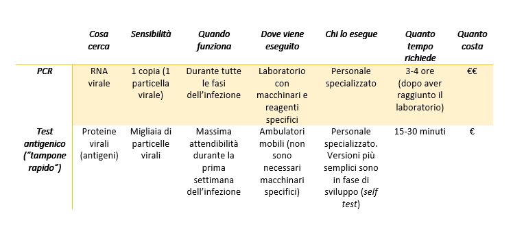
In the case of the swab tests we have heard since the beginning of the pandemics, the aim is to detect the genetic material of the virus.

  • The viral RNA is extracted from the sample (don’t forget the genome of SARS-CoV-2 is made of RNA, not DNA)
  • The RNA is “transformed” into DNA through a reaction called reverse transcription
  • The DNA is in turn copied several times in a PCR (polymerase chain reaction), a technique able to detect even very small quantities of DNA.
  • If the patients from which the sample has been collected does not have the virus, there is no DNA to copy, and the result of the PCR will be negative. On the other hand, if in the sample is present even only one copy of viral RNA, the reverse transcription and the PCR will be able to recognize and amplify it, and the result will be positive.

When the proteins of the virus (antigens) are the target of the tests, we talk about rapid swab tests (antigenic tests). The working principle is very similar to that of the serologic tests (see this post), but in this case, specific antibodies against the proteins of SARS-CoV-2 are immobilized on the device.

  • The material collected with the swab is placed in a well at one extremity of the device
  • The sample will flow by capillarity through the strip and it will go across the conjugation chamber, where it will encounter antibodies specific for the viral proteins conjugated to colloidal gold (pink-red coloured)
  • If the sample contains viral antigens, they will bind the antibodies and will carry them while flowing in the strip until they encounter other antibodies against the same proteins, bound to the device
  • The viral proteins will be trapped in a “sandwich” between the antibodies immobilized on the device and those conjugated to the colloidal gold particles, and this will determine the appearance of a red band, revealing the presence or absence
  • The intensity of the band can indicate whether the amount of viral proteins in the sample is more or less abundant.

(The antigenic tests, similarly to the serologic ones, have an internal control to ensure the validity of the result).

While the PCR can detect very small amounts of genetic material, and therefore can be used in any phase of the infection, including on asymptomatic patients, the sensitivity of the antigenic tests is lower (the result is positive only if the sample contains a large amount of proteins). For this reason, the antigenic tests are used only on symptomatic patients to determine the cause of their symptoms, but they are not an official method for the screening of the virus: the results from antigenic tests need to be confirmed with a PCR, that is for the time being the only official diagnostic method.

Both the PCR and the antigenic tests must be done by healthcare professionals; there are no approved tests for the self-diagnosis, even if some companies are working on a simplified version of the rapid tests to be used at home, at school on at workplaces (the first data on the saliva-based antigenic test are not satisfactory).

Comparison between PCR and antigenic tests

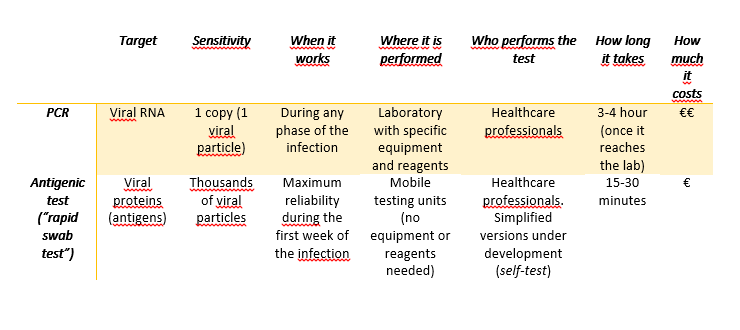
Image created in BioRender.com by Carla Usai

Bibliography

Fast Coronavirus tests are coming, Guglielmi G., Nature 2020 https://www.nature.com/articles/d41586-020-02661-2

European Centre for Disease Prevention and Control: https://www.ecdc.europa.eu/sites/default/files/documents/Overview-rapid-test-situation-for-COVID-19-diagnosis-EU-EEA.pdf

World Health Organization: https://www.who.int/publications/i/item/antigen-detection-in-the-diagnosis-of-sars-cov-2infection-using-rapid-immunoassays

Come funzionano i tamponi (rapidi e non) per SARS-CoV-2

Se un anno fa mi avessero detto che le nostre conversazioni quotidiane avrebbero avuto come parole chiave “virus, PCR, tampone, anticorpi” non ci avrei mai creduto, e invece (ahinoi!) questi argomenti stanno dominando il 2020.

Visto che tutti ne parlano, ma molti fanno ancora confusione, ecco qui le differenze tra “tamponi” e “tamponi rapidi” (come vengono chiamati colloquialmente).

Il tampone in sé e per sé non è altro che il bastoncino di cotone con cui viene raccolto il materiale presente nella cavità nasale o oro-faringea.

La differenza sta nel modo in cui il materiale prelevato viene analizzato.

Nel caso dei tamponi di cui abbiamo sentito parlare dall’inizio della pandemia viene ricercato il materiale genetico del virus:

  • Dal campione prelevato con il tampone viene estratto l’RNA (ricordiamo che il genoma di SARS-CoV-2 non è fatto di DNA bensì di RNA)
  • L’RNA che viene “trasformato” in DNA in laboratorio mediante una reazione chiamata retro trascrizione
  • Il DNA così ottenuto viene copiato moltissime volte tramite PCR (reazione a catena della polimerasi), che ci consente di rilevare anche quantità molto piccole di DNA.
  • Se il paziente da cui è stato prelevato il campione non ha il virus, non ci sarà nessun DNA da copiare e il risultato sarà negativo. Ma se nel campione è presente anche una sola copia di RNA virale, la retro trascrizione e la PCR saranno in grado di riconoscerla e amplificarla, e il risultato del test sarà positivo.

Quando si parla di tamponi rapidi ci si riferisce invece alla ricerca nel campione delle proteine del virus (antigeni). Il funzionamento è simile a quello dei test sierologici (vedi questo post), ma in questo caso sulla membrana del dispositivo sono immobilizzati degli anticorpi specifici contro le proteine di SARS-CoV-2:

  • Ad una estremità del dispositivo è presente un pozzetto in cui viene deposto il materiale prelevato con il tampone
  • Il campione inizierà a muoversi per capillarità lungo la striscia di resina, e incontrerà in una porzione di essa (la camera di coniugazione) degli anticorpi specifici per proteine del virus coniugati a delle particelle d’oro colloidale che hanno un colore rosa-rossastro.
  • Se nel campione sono presenti gli antigeni virali, questi si uniranno agli anticorpi e li trascineranno con sé mentre continuano a muoversi per capillarità lungo la striscia fino a quando non incontreranno degli altri anticorpi contro gli stessi antigeni, immobilizzati sul dispositivo.
  • Gli antigeni del virus si troveranno così intrappolate in un “sandwich” tra gli anticorpi immobilizzati sulla resina e quelli legati all’oro colloidale, e ciò determinerà la comparsa di una banda rossa, rendendo facilmente visibile la loro presenza o assenza.
  • L’intensità del colore della banda potrà indicarci se le proteine del virus presenti nel tampone sono più o meno abbondanti.

(Anche i test antigenici, come quelli sierologici, sono dotati di un controllo interno per assicurare la validità del risultato).

Mentre la PCR è in grado di individuare anche quantità molto piccole di materiale genetico, e quindi può essere usata in tutte le fasi dell’infezione, incluso sui pazienti asintomatici, la sensibilità dei test antigenici è decisamente inferiore (il risultato è positivo solo se è presente una quantità notevole di proteine). Per questo al momento i test antigenici vengono impiegati solo su pazienti sintomatici per confermare la causa dei sintomi, ma non sono un metodo ufficiale per lo screening: il risultato dei test antigenici deve essere confermato tramite PCR, che rimane l’unico metodo diagnostico ufficiale.

Sia la PCR che il test antigenico devono essere eseguiti da personale specializzato; non esistono ancora test autorizzati per l’auto diagnosi, anche se si sta cercando di sviluppare versioni più semplici dei test rapidi che possano essere eseguiti autonomamente a casa, nelle scuole o nei luoghi di lavoro (i primi dati del test antigenico salivare non sono però del tutto soddisfacenti).

Le caratteristiche principali di PCR e test antigenici a confronto.

Immagine creata con BioRender.com da Carla Usai

Bibliografia

Fast Coronavirus tests are coming, Guglielmi G., Nature 2020 https://www.nature.com/articles/d41586-020-02661-2

European Centre for Disease Prevention and Control: https://www.ecdc.europa.eu/sites/default/files/documents/Overview-rapid-test-situation-for-COVID-19-diagnosis-EU-EEA.pdf

Organizzazione Mondiale della Sanità: https://www.who.int/publications/i/item/antigen-detection-in-the-diagnosis-of-sars-cov-2infection-using-rapid-immunoassays

Il Virus del Nilo Occidentale (West Nile Virus)

Nota dell’autrice: questo articolo è stato pubblicato per la prima volta il 15/09/2020; ultimo aggiornamento: 28/07/2025.

Che cos’è il Virus del Nilo Occidentale e dove è diffuso?

Il Virus del Nilo Occidentale, o West Nile Virus (WNV), nonostante abbia un nome esotico, è ormai di casa anche in Europa. Nel 2025 sono stati riportati casi in Grecia, Romania e Italia, con casi recenti in Piemonte, Emilia Romagna, Lazio e Veneto.

Inizialmente era presente in Africa (fu isolato nel 1937 in Uganda nella regione del Nilo Occidentale), da cui si diffuse in parte dell’Europa, Medio Oriente, Asia Occidentale e Australia, seguendo le rotte degli uccelli migratori. Nel 1999 raggiunse New York e da allora si è esteso a tutto il Nord America e in Venezuela, diventando l’arbovirus più diffuso al mondo. La diffusione del WNV in nuove aree è stata facilitata dal trasporto (non intenzionale) di insetti vettori con navi e aerei, da attività umane come urbanizzazione e irrigazione dei campi, e da cambiamenti climatici (variazioni di temperatura e precipitazioni atmosferiche) che hanno favorito la proliferazione delle zanzare.

In quale periodo dell’anno si diffonde il virus del Nilo Occidentale?

La diffusione del virus avviene quando le zanzare sono attive (primavera-estate), e di conseguenza i casi di infezione nell’uomo e nei cavalli nell’emisfero Boreale vengono registrate prevalentemente tra luglio e settembre.

Come si trasmette il virus del Nilo Occidentale?

Il virus West Nile è un arbovirus (arthropode borne virus) trasmesso dalla zanzara comune (Culex pipiens). Il virus circola trasportato dalle zanzare (che ne sono i vettori) e montiplicandosi negli uccelli selvatici che ne costituiscono il serbatoio. Questa modalità di circolazione è detta ciclo enzootico.

Il virus viene trasmesso agli uccelli selvatici tramite punture di zanzara, e occasionalmente può infettare equini e uomo (più raramente anche altri mammiferi) se questi vengono punti da una zanzara portatrice del virus: si tratta quindi di un virus zoonotico, cioè trasmesso dagli animali all’uomo. Questo virus non sembra causare nessuna malattia negli insetti.

Gli uccelli vengono infettati facilmente dal virus West Nile e ne sono il serbatoio naturale consentendone un’efficiente replicazione, mentre equini e umani sono considerati ospiti occasionali; questi ultimi non contribuiscono alla diffusione del virus in quanto il numero di particelle virali nel sangue (viremia) resta basso. Negli ultimi 20 anni è stato registrato un aumento delle infezioni negli ospiti occasionali.

Il ciclo di trasmissione del Virus del Nilo Occidentale. Immagine creata su BioRender.com da Carla Usai

Come fanno le zanzare a trasmettere il virus West Nile?

Le zanzare si infettano nutrendosi del sangue di un volatile infetto. Il virus raggiunge così le ghiandole salivari della zanzara, e può essere successivamente iniettato in un altro ospite, dove può moltiplicarsi e in alcuni casi causare la malattia.

Per nutrirsi del sangue di un vertebrato, le zanzare iniettano nei vasi sanguigni della vittima un po’ di saliva che, oltre ad avere proprietà anticoagulanti, contiene delle sostanze che inibiscono alcune cellule del sistema immunitario (i linfociti T), facilitando così la diffusione del virus.

Le persone infettate dal virus West Nile sono contagiose?

Gli esseri umani che hanno contratto WNV non sono direttamente contagiosi, ma il virus può essere trasmesso tramite trasfusioni di sangue o trapianto di organi (i donatori di sangue vengono testati per la presenza del virus e non è permessa la donazione nei primi 28 giorni al ritorno da zone in cui il virus è endemico). È stato registrato un solo caso di trasmissione da madre a figlio durante la gravidanza e un solo caso di contagio tramite latte materno.

Esiste invece la possibilità di contagio diretto tra volatili senza l’intervento di insetti vettori.

Quali cellule vengono colpite dal virus West Nile?

Inizialmente il WNV infetta le cellule dell’epidermide (cheratinociti), e le cellule del sistema immunitario associate alla pelle (macrofagi e cellule di Langerhans). Mentre i macrofagi sono in grado di eliminare il virus, le cellule di Langerhans infettate lo trasportano ai linfonodi, da cui può diffondersi ad altri organi e infettare altri tipi di cellule.

Che malattia causa il virus West Nile?

La maggior parte delle infezioni nell’uomo è asintomatica, mentre circa il 20% delle infezioni causa la Febbre del Nilo Occidentale (West Nile FeverWNF). In meno dell’1% dei casi il virus raggiunge le cellule del Sistema Nervoso Centrale causando la Malattia Neuroinvasiva del Nilo Occidentale (West Nile Neuroinvasive Disease, WNND); ciò avviene solitamente in individui anziani, immunocompromessi, o debilitati da altre patologie.

Quali sono i sintomi di un’infezione da virus West Nile?

La Febbre del Nilo Occidentale (WNF) è caratterizzata dall’improvvisa comparsa di sintomi come mal di testa, malessere, dolori muscolari e agli occhi, vomito, febbre, itterizia (colorazione gialla di pelle, occhi e mucose)  ed eruzioni cutanee. Di solito i sintomi passano nell’arco di una settimana, ma in alcuni casi possono durare fino ad alcuni mesi e richiedere il ricovero.

La Malattia Neuroinvasiva del Nilo Occidentale (WNND) invece, può causare meningite, encefalite, paralisi flaccida o una combinazione di queste manifestazioni, e portare a morte nel 17% dei casi.

Anche nei cavalli l’infezione è solitamente asintomatica, ma nel 10% dei casi gli animali mostrano difficoltà nei movimenti fino alla totale incapacità di sollevarsi sulle zampe, fino al sopraggiungere della morte.

Come ci si difende dal virus West Nile?

Al momento non esiste un vaccino ad uso umano (ma è disponibile un vaccino per i cavalli), e non esiste una cura specifica per contrastare questa infezione.

Per tenere sotto controllo la propagazione del virus ed evitare l’infezione, è possibile agire su più fronti:

  • screening dei donatori di sangue e organi per evitare la trasmissione tramite sangue e organi infetti
  • monitoraggio degli animali serbatoio per individuare la presenza del virus sul territorio
  • riduzione delle zone di acque stagnanti che ospitano le larve delle zanzare
  • uso di repellenti e zanzariere per ridurre la possibilità di entrare in contatto con zanzare adulte infette.

Come è fatto il Virus del Nilo Occidentale?

La particella virale è di sferica con un diametro di circa 50 nanometri (= 50 miliardesimi di metro, o 50 milionesimi di millimetro). WNV appartiene alla famiglia dei Flaviviridae, come il virus dell’epatite C (HCV) e il virus Zika; il suo genoma a RNA codifica per 10 proteine ed è racchiuso in un rivestimento proteico (capside) circondato a sua volta da una membrana lipidica.

Esistono 8 tipi diversi di WNV (genotipi) esistenti in natura, ma i genotipi 1 e 2 causano una malattia nell’uomo.

Bibliografia

Habarugira G. West Nile Virus: An Update on Pathobiology, Epidemiology, Diagnostics, Control and “One Health” Implications. Pathogens. 2020. https://www.mdpi.com/2076-0817/9/7/589

Istituto Superiore di Sanità (ISS). Febbre West Nile – News. Epicentro. Consultato il 28 luglio 2025. https://www.epicentro.iss.it/westnile/aggiornamenti

World Health Organisation. West Nile virus. Consultato il 28 luglio 2025. https://www.who.int/news-room/fact-sheets/detail/west-nile-virus

European Centre for Disease Control and Prevention. Factsheet about West Nile virus infection. Consultato il 28 luglio 2025. https://www.ecdc.europa.eu/en/west-nile-fever/facts

European Centre for Disease Control and Prevention. Weekly updates: 2025 West Nile virus transmission season. July 28, 2025. Consultato il 28 luglio 2025. https://www.ecdc.europa.eu/en/west-nile-fever/surveillance-and-disease-data/disease-data-ecdc

West Nile Virus (WNV)

Author’s note: this article was published for the first time on 15/09/2020; latest update: 28/07/2025

What is the West Nile Virus and where is it found?

West Nile Virus (WNV), despite its exotic name, is quite a common virus in Europe. In 2025 cases have been reported in Greece, Romania and Italy.

It was initially present in Africa (when it was isolated for the first time in 1937 in the West Nile region in Uganda), from where it spread to of Europe, the Middle East, Western Asia, and Australia, along the routes of migratory birds.In 1999 the virus arrived in New York and spread to all North America and Venezuela, being now the arbovirus with the widest geographical distribution. The spread of WNV to new areas has been made easier by the unintentional transportation of vector mosquitoes with ships and airplanes, by human activities such as urbanization and field irrigation, and by climate changes (higher temperatures and different precipitation patterns) that have promoted the proliferation of vector mosquitoes.

Does the West Nile virus spread in certain times of the year?

The virus can spread when mosquitoes are active (spring-summer), therefore infections in humans and horses in the Northern hemisphere are mainly reported from July to September.

How does WNV spread?

WNV is an arbovirus: a virus transmitted by arthropods (arthropod-borne virus), in particular by the common mosquitoes (Culex pipiens), and it is maintained in nature in a transmission cycle involving mosquitoes (as vectors) and wild birds (as a reservoir). This type of transmission is called enzootic cycle.

The virus is transmitted to wild birds through mosquito bites, and it can occasionally infect humans and horses (more infrequently also other mammals) when they are bitten by a mosquito carrying the virus: it is therefore a zoonotic virus, transmitted from animals to humans. WNV is thought not to cause any disease in insects.

Birds are the specific host and natural reservoir for WNV, supporting an efficient viral replication, while humans and horses are occasional hosts that do not contribute to the spread of the virus, since the number of virual particles in their blood (viremia) remains low. There has been an increase in the infection rate in these two species for the last 20 years.

How do mosquitoes spread WNV?

Mosquitoes acquire the virus through blood meals on infected birds. The virus migrates to the salivary glands and can be later injected into another host where it can replicate and eventually cause disease. To feed on the blood of a vertebrate, mosquitoes inject into the blood vessels of their victims a small quantity of saliva, that, besides avoiding clotting, inhibits the activity of an immune cell type (T cells), allowing the spread of the virus.

Transmission cycle of the West Nile Virus. Created in BioRender.com by Carla Usai

Is West Nile Virus contagious?

People infected by WNV are not directly contagious, but the virus can be transmitted to other individuals through blood transfusion or organ transplantation (donors are screened for the presence of WNV –among other viruses- and donation is not allowed within 28 days after arrival from WNV endemic areas). A single case of mother-to-child transmission during pregnancy, as well as a single case of contagion through breastfeeding, have been reported so far.

On the contrary, the virus can be directly transmitted among birds even without vector involvement.

What cells does WNV infect?

Initially, WNV infects the cells of the epidermis (keratinocytes), and some immune cells located in the skin (macrophages and Langerhans cells). While macrophages can rapidly eliminate the virus, infected Langerhans cells migrate to the lymph nodes, from where the virus can spread to other organs and infect a wide range of cell types.

Does WNV cause a disease?

Most of the infections in humans are asymptomatic, but in about 20% of cases, it causes a disease called West Nile Fever (WNF). In less than 1% of cases, WNV leads to the more severe West Nile Neuroinvasive Disease (WNND) when cells of the central nervous system are infected, usually in elderly patients, immunocompromised or affected by other pathologies.

What are the symptoms of WNV infection?

West Nile Fever (WNF) is characterised by the sudden appearance of symptoms such as headache, malaise, muscle soreness, eye pain, nausea, fever, jaundice, and rash. People usually start to feel better in about a week, but sometimes symptoms last for months and require hospitalisation.

West Nile Neuroinvasive Disease (WNND) causes meningitis, encephalitis, flaccid paralysis, or a combination of them, and it leads to death in 17% of cases.

In horses as well, the infection is usually asymptomatic, but in 10% it causes movement disorders, including the inability to stand up, and death.

How can we prevent WNV spread?

Unfortunately, there is no WNV vaccine for humans (but there is one for horses) and to date, there is no specific treatment either.

Nevertheless, the spreading of the virus can be controlled by:

  • screening of blood and organ donors
  • monitoring of reservoir populations for early detection of the virus in the area
  • reduction of standing water areas able to host mosquito larvae
  • use of repellents and mosquito nets per reduce exposure to infected adult mosquitoes.

What does WNV look like?

The viral particle is sphere-shaped, with a diameter of about 50 nanometers (= 50 billionths of a meter, or 50 millionths of a millimeter).  WNV is an RNA virus belonging to the Flaviviridae family, which includes the Hepatitis C virus (HCV) and Zika virus as well; its genome encodes 10 different proteins and is surrounded by both a proteic capsid and a lipidic envelope.

There are 8 different types of WNV (genotypes), but only genotypes 1 and 2 cause disease in humans.

Bibliography

Habarugira G. West Nile Virus: An Update on Pathobiology, Epidemiology, Diagnostics, Control and “One Health” Implications. Pathogens. 2020. https://www.mdpi.com/2076-0817/9/7/589

Istituto Superiore di Sanità (ISS). Febbre West Nile – News. Epicentro. Accessed 28/07/2025. https://www.epicentro.iss.it/westnile/aggiornamenti

World Health Organisation. West Nile virus. Accessed 28/07/2025. https://www.who.int/news-room/fact-sheets/detail/west-nile-virus

European Centre for Disease Control and Prevention. Factsheet about West Nile virus infection. Accessed 28/07/2025. https://www.ecdc.europa.eu/en/west-nile-fever/facts

European Centre for Disease Control and Prevention. Weekly updates: 2025 West Nile virus transmission season. July 28, 2025. Accessed 28/07/2025. https://www.ecdc.europa.eu/en/west-nile-fever/surveillance-and-disease-data/disease-data-ecdc

Per aspera ad astra: l’importanza dei fallimenti nella scienza (ma non solo)

Nicolás Espinosa / CC BY-SA (https://creativecommons.org/licenses/by-sa/4.0)

Come scienziati siamo abituati all’idea del fallimento.

Gli esperimenti che non funzionano sono più di quelli che vanno esattamente come previsto, i papers che subiscono revisioni e correzioni sono enormemente più numerosi di quelli che vengono pubblicati al primo tentativo senza nessuna modifica, le richieste di finanziamento respinte superano quelle concesse. La mole di lavoro e il tempo necessari per iniziare a vederne i risultati possono essere talvolta sconfortanti.

Tuttavia di fallimenti si parla poco. Eppure i fallimenti, le sconfitte e le delusioni in senso lato sono il nostro pane quotidiano, fanno parte della vita del ricercatore, sono anch’essi, se vogliamo, quei tentativi ed errori alla base del metodo scientifico.

Siamo più propensi a celebrare le vittorie che a riconoscere le sconfitte. Presentiamo solo i risultati positivi e le ipotesi confermate, quasi mai quelle smentite.*

Recentemente sono stata invitata a partecipare come relatrice ad una giornata formativa per ragazze interessate a intraprendere studi scientifici. L’evento, STEMinist,  è stato organizzato da una dottoranda della Queen Mary University of London (che potete trovare su Twitter come @Sciencythoughts) e patrocinato dal Centre for Public Engagement della stessa università, con lo scopo di fornire esempi di donne che ricoprono diversi ruoli in ambito scientifico a ragazze arrivate in UK come rifugiate, condividere con loro le nostre storie e dimostrare che, indipendentemente dalla nostra (variegata) provenienza sociale, tutte siamo riuscite a raggiungere i nostri obiettivi.

Il mio intervento era il primo in programma; l’organizzatrice mi aveva chiesto di parlare della mie esperienze nei diversi paesi europei in cui ho studiato e lavorato, e di dare un’idea del mio ambito di ricerca, ma non avevo idea di come sarebbero stati gli altri interventi, e a dire il vero temevo di dire qualcosa di banale.

Ma mentre preparavo la presentazione ripercorrendo questi ultimi 15 anni (!) ho finalmente avuto una visione di insieme del mio percorso: mi sono resa conto per la prima volta degli obiettivi raggiunti, delle difficoltà superate, delle soddisfazioni e delle delusioni. Insomma, mi sono resa conto di come le sconfitte che ho collezionato via via mi abbiano portato in giro per il mondo fino a farmi atterrare a Londra!

Se avessi ottenuto una borsa di dottorato subito dopo la Laurea non avrei mai frequentato il Centro Nacional de Biotecnologia di Madrid con il progetto Leonardo Da Vinci; se avessi avuto la possibilità di restare a Madrid non avrei partecipato a quel congresso a Seul in cui mi sono confrontata a tu per tu con scienziati illustri. Se non avessi rinunciato a quel primo lavoro che non mi soddisfaceva, non avrei mai costruito la rete di contatti che ho adesso e che ha fatto sì che mi venisse offerto un contratto a Londra.

Il messaggio che ho voluto trasmettere a il nostro giovane pubblico è stato di credere sempre nelle seconde possibilità e di non paragonare mai il proprio percorso a quello degli altri, ma soprattutto di considerare che da ogni esperienza, positiva o negativa che sia, possiamo imparare qualcosa.

Dopo di me è stato il turno di altre scienziate, ognuna in una tappa diversa della propria carriera scientifica, e mi ha piacevolmente colpito il fatto che ognuna di noi abbia parlato dei propri fallimenti e delusioni come passaggi fondamentali per raggiungere i nostri obiettivi.

Tutte le relatrici hanno raccontato di essersi trovate in un momento o in un altro davanti ad un bivio e di aver avuto dei dubbi sulla strada da intraprendere; molte di noi hanno avuto un inizio di carriera deludente con un lavoro che non è risultato come ci aspettavamo o con un’opportunità sfumata; qualcuna ha parlato di aver affrontato ansia e depressione, altre delle difficoltà di iniziare un dottorato avendo una famiglia da gestire, quasi tutte siamo state le prime nelle nostre rispettive famiglie a intraprendere una carriera accademica con tutti gli svantaggi che questa situazione può comportare.

Allora forse non sarebbe il caso di normalizzare i fallimenti? Di educare all’insuccesso e a imparare da esso invece di nasconderlo come un’onta?

Sul tema sono stati pubblicati libri (Failure: Why Science Is So Successful, di Stuart Firestein), a Modena è stata fondata una vera e propria scuola di fallimento dedicata a giovani imprenditori, studenti e disoccupati (www.scuoladifallimento.com), e addirittura recentemente è stato sviluppato un modello matematico che descrive con un’equazione quali tentativi infruttuosi porteranno a futuri risultati positivi.

E c’è anche chi, come Brian W. Jones della Univeristy of Utah, e Johannes Haushofer, professore a Princeton, hanno reso pubblica una cronologia dei propri fallimenti.

Al di là delle teorie e delle equazioni, voglio condividere con voi tre messaggi importantissimi che sono emersi più volte nel corso di STEMinist, e che forse possono aiutarci dal punto di vista pratico a considerare successi e fallimenti nella giusta prospettiva:

  • Non esiste un’unica strada verso il successo: ognuno di noi segue il proprio personalissimo percorso.
  • Tutte le esperienze sono formative: ogni cosa che facciamo ci avvicina alla meta, anche se spesso non ce ne accorgiamo.
  • Dobbiamo imparare a conoscerci ed essere fedeli a noi stessi: l’idea di successo da perseguire è diversa per ciascuno di noi, e in fin dei conti le uniche aspettative da soddisfare sono le nostre.

Buon insuccesso a tutti e ad maiora!

*(N.B. A mio modesto parere, se comunicare i risultati negativi fosse prassi nella comunicazione scientifica, forse non ci troveremmo in situazioni come quella attuale in cui il pubblico esige che gli scienziati trovino immediatamente una cura per una malattia appena scoperta, o peggio ancora non ci sentiremmo dire che la cura per il cancro esiste ma viene tenuta nascosta.)

Bibliografia

Failure: Why Science Is So Successful, Stuart Firestein, Oxford University Press, 2015

A celebration of failure, Loscalzo J., Circulation 2014, http://doi.org/10.1161/CIRCULATIONAHA.114.009220

Quantifying the dynamics of failure across science, startups and security, Yin Y. et al., Nature 2019, https://doi.org/10.1038/s41586-019-1725-y

Scientific progress is built on failure, Parkes E., Nature career 2019 http://doi.org/10.1038/d41586-019-00107-y

Curriculum vitae dei fallimenti, Johannes Haushofer https://www.princeton.edu/~joha/Johannes_Haushofer_CV_of_Failures.pdf

Cronologia delle richieste di finanziamento vinte e di quelle respinte, Bryan W. Jones https://twitter.com/BWJones/status/1143616543470710784

Per aspera ad astra: failure and success in (and beyond) science

Nicolás Espinosa / CC BY-SA (https://creativecommons.org/licenses/by-sa/4.0)

As scientists, we are familiar with the concept of failure.

The experiments that do not work are more than those that actually do, the papers that need revisions and corrections exceed those published without any correction, the number of rejected grant applications is greatly larger than the number of applications that are accepted. The effort and the time necessary to start seeing the result of our work can sometimes be disheartening

However, we rarely talk about failure. Nevertheless, failure and disappointment form a scientist’s daily routine, and they are part of that trial-and-error process at the basis of the scientific method.

We are more inclined to celebrate our victories than to admit the defeat. We do not share our failed attempts on our social media profiles. We only present positive results and verified hypothesis, rarely the rejected ones.*

Recently, I have participated as a speaker in an educational meeting for girls interested in pursuing a scientific career. The event, STEMinist, was organised by a Ph.D. student at Queen Mary University of London (she is on Twitter as @Sciencythoughts) and funded by the Centre for Public Engagement of the same university, to present women with different roles in science to young refugee women, share our experience with them, and show them that regardless our most diverse social and cultural backgrounds, we all have achieved our goals.

My talk was scheduled to be the first one in the morning; the organiser had asked me to talk about my experience in the different European countries in which I had studied and worked and to briefly introduce my research topic. But I had no idea of what the other talks would have looked like, and, to be honest, I was afraid to fall into triteness.

But when I started to recall the last 15 years (!) to prepare my presentation, I finally got the overall view of my career path: I realized perhaps for the first time, the achievements, the difficulties, the gratifications, and the failures. In other words, I realized that all the defeats that I have collected have brought me around the world all the way to London!

Had I obtained a scholarship right after my Master’s degree, I would have never worked in the Spanish National Centre for Biotechnology with a Leonardo da Vinci fellowship; had I had the possibility to stay in Madrid longer, I would have never attended that congress in Seoul where I met all those renowned scientists. Hadn’t I given up that first job that I did not like, I would have never built my network that in the end resulted in a job offer in London.

The message that I wanted to convey to our young audience was to always believe in second chances, to never compare ourselves to others, and, most importantly, to consider every experience as a learning experience.

I am glad that all the other speakers, each one in a different phase of their scientific career, also talked about their struggles and failures as instrumental to achieve their goals.

Each and every speaker talk about having had, at some point, doubts about the path to follow; most of us had a disappointing first job or a rejection at the beginning of our career; some talk about anxiety and depression, other about the struggles of parenting and embarking on a doctorate at the same time; almost all of us have been first-generation Ph.D. students, with all the disadvantages that it implies.

So why not we try to normalize failure? To educate to failure and learn from it, instead of hiding it as something to be ashamed of?

There are books on this topic (like Failure: Why Science Is So Successful, by Stuart Firestein), in Modena (Italy) there is an actual school of failure addressed to young intrapreneurs, students and unemployed people (www.scuoladifallimento.com), and even a mathematical model has been developed to describe with an equation what failed attempts would lead to successful results.

What is more, Brian W. Jones of the University of Utah, and Johannes Haushofer, professor at Princeton, have released a timeline of their failures!

All these theories and equations aside, I would like to share with you three very important messages that emerged repeatedly during STEMinist, and I hope could help us from a particle point of view to put failure and success into the right perspective:

  • There is no set path to success: everyone is in their own very personal journey
  • Every experience is a learning experience: what you are doing will take you places (even if you don’t know yet)
  • We need to know ourselves and be true to our hopes: the idea of success can be very different for each person, and in the end, the only expectations worth fulfilling are our own.

Embrace your failures and ad maiora!

*(In my humble opinion, if negative results were regularly communicated in science, we may not be now in a situation where the general public demands treatments for a newly discovered disease, or, what is worse,  there would not be so many people claiming that the cure for “cancer” exists but is kept hidden.)

Bibliography

A celebration of failure, Loscalzo J., Circulation 2014, http://doi.org/10.1161/CIRCULATIONAHA.114.009220

Quantifying the dynamics of failure across science, startups and security, Yin Y. et al., Nature 2019, https://doi.org/10.1038/s41586-019-1725-y

Scientific progress is built on failure, Parkes E., Nature career 2019 http://doi.org/10.1038/d41586-019-00107-y

CV of failures, Johannes Haushofer https://www.princeton.edu/~joha/Johannes_Haushofer_CV_of_Failures.pdf

Timeline of rejected and successful grant applications, Bryan W. Jones https://twitter.com/BWJones/status/1143616543470710784

Failure: Why Science Is So Successful, Stuart Firestein, Oxford University Press, 2015

I virus del passato intrappolati nei ghiacciai

Già dagli inizi del secolo passato è noto che i ghiacciai conservano nelle loro profondità microorganismi intrappolati da migliaia di anni.

Lo studio di campioni di ghiaccio prelevati tramite carotaggio ha consentito di risalire a le condizioni climatiche che si sono succedute nel tempo, in base alle caratteristiche dei batteri in essi contenuti. In alcuni casi sono stati trovati dei batteri ancora vivi di cui è stato possibile riattivare il metabolismo per comprendere di che sostanze si nutrissero, che sostanze di scarto producessero, e quali fossero le condizioni ambientali migliori per la loro sopravvivenza.

Mentre molti studi sono stati condotti sulle popolazioni batteriche intrappolate nei ghiacciai, le informazioni riguardo i virus sono ancora scarse. Questo è dovuto alla bassissima concentrazione di particelle virali nei ghiacciai (circa 1000 volte inferiore alla concentrazione in cui si trovano nell’acqua di mare) e alle maggiori difficoltà tecniche per individuare acidi nucleici virali rispetto al DNA batterico.

Recentemente un gruppo di ricercatori della Ohio State University, University of Nebraska e Lawrence Berkeley National Laboratory, è riuscito a isolare batteri e virus diversi da due campioni prelevati a diverse profondità dal ghiacciaio Guliya (Tibet, Cina). Utilizzando nuove tecniche di sequenziamento sono riusciti a ricostruire 33 sequenze di virus rimasti intrappolati circa 500 e 15000 anni fa; il confronto delle sequenze ottenute con le sequenze di 2304 virus conosciuti, ne ha permesso una parziale classificazione.

Il materiale genetico ritrovato appartiene a diverse popolazioni di batteriofagi, ossia di virus che infettano i batteri. Tramite analisi bioinformatiche gli scienziati sono riusciti ad predire quali fossero i batteri target di questi virus. La frequenza dei diversi tipi di batteriofagi trovati nei due campioni delle due diverse epoche corrisponde alla distribuzione delle specie batteriche presenti: in ciascuno dei due campioni i batteri più abbondanti costituivano proprio le popolazioni target predette per i batteriofagi più abbondanti.

Anche se per la metà dei virus isolati non è stato possibile identificare i batteri target, i risultati di questo studio confermano che i virus, così come altre condizioni ambientali quali temperatura, radiazioni, concentrazioni di gas e altre sostanze chimiche, svolgono un ruolo molto importante nel modellare gli ecosistemi.

Ricotruzione 3D di un batteriofag, di Reo Kometani and Sunao Ishihara / CC BY (https://creativecommons.org/licenses/by/4.0)

Bibliografia:

Glacier ice archives fifteen-thousand-year-old viruses,  Zhong Z-P. et al., bioRxiv preprint 2020 https://doi.org/10.1101/2020.01.03.894675

Viruses from the past trapped in glacier ice

It has been known since the beginning of the last century, that glaciers store in their depths microorganisms from thousands of years ago.

The study of ice samples collected using core drills has allowed determining the environmental conditions of different ages, based on the characteristics of the bacteria they contain. In some cases, bacteria found in ice samples were still alive, and it was possible to reactivate their metabolism to understand which nutrients they used, which molecules they produced, and in which optimal conditions they lived.

While several studies have been done about bacteria trapped in the glaciers, information about viruses is still scarce. This is due both to the very low concentration of viral particles in this kind of sample (about 1000 fold lower than in seawater) and to the fact that is technically more difficult to identify viral nucleic acids than bacterial DNA.

Recently, a group of researchers from Ohio State University, University of Nebraska, and  Lawrence Berkeley National Laboratory has isolated bacteria and viruses from two ice samples collected at two different depths in the Guliya ice cap (Tibet, China). Using modern sequencing techniques, the researchers were able to determine 33 different viral sequences trapped in the ice 500 and 15000 years ago; comparison with the sequences of 2304 known virus, has allowed a partial classification.

The genetic material found in the samples belongs to different populations of bacteriophages, viruses that infect bacteria, and the bacterial target populations of some of them were predicted through bioinformatics analyses. The frequency of the different types of bacteriophages found in the two samples from different ages correlates with the distribution of the bacterial species: in each of the two samples, the most abundant bacteria are indeed the target populations predicted for the dominant bacteriophages.

Even if it was not possible to determine the target bacteria for half of the isolated viruses, the results of this study confirm that viruses, as well as other environmental conditions such as temperature, radiations, and chemical concentrations, play an important role in shaping the ecosystems.

3D reconstruction of a bacteriophage by Reo Kometani and Sunao Ishihara / CC BY (https://creativecommons.org/licenses/by/4.0)

Bibliography:

Glacier ice archives fifteen-thousand-year-old viruses,  Zhong Z-P. et al., bioRxiv preprint 2020 https://doi.org/10.1101/2020.01.03.894675

I virus come alleati: la terapia genica per la distrofia retinica e la beta-talassemia.

La terapia genica si pone l’obiettivo di curare le malattie genetiche monofattoriali, cioè quelle causate da mutazioni all’interno di un singolo gene.

Le sequenze di basi nel DNA vengono “lette” in gruppi di tre (triplette o codoni) per ottenere le istruzioni per costruire le proteine, e ogni tripletta di basi corrisponde a un amminoacido. Qualsiasi alterazione all’interno della sequenza di DNA (mutazione) si ripercuote sulla sequenza di amminoacidi e quindi sulla funzione della proteina.

Anche mutazioni a carico di una singola base azotata possono avere conseguenze devastanti sulla proteina finale. Per esempio la sostituzione di una base con una differente può far cambiare l’amminoacido codificato da quella tripletta (mutazione missenso), mentre l’inserzione o la perdita (delezione) di una base fanno slittare la successione delle triplette causando la traduzione di una sequenza di amminoacidi totalmente diversa (mutazione frameshift).

In alcuni casi la sostituzione di una base può trasformare la tripletta originale in un codone di stop, ovvero una tripletta che indica la fine della proteina (mutazione non-senso), questo tipo di mutazioni causa una interruzione prematura della sequenza di amminoacidi dando vita a una proteina tronca che non sarà in grado di svolgere la sua funzione.

Talvolta la sostituzione di una base trasforma il codone iniziale in una tripletta che codifica per lo stesso amminoacido; mutazioni di questo tipo non hanno nessun effetto sulla sequenza amminoacidica della proteina e vengono dette mutazioni silenti.

Se a causa di una mutazione in un gene, la proteina che viene prodotta non è capace di svolgere correttamente la sua funzione, il risultato è una malattia. Per annullare gli effetti di mutazioni che interessano una singola proteina, si può fornire alle cellule una copia corretta del gene che la codifica.

Per far arrivare un frammento di DNA alle cellule che ne hanno bisogno è necessario usare qualcosa che possa trasportarlo, un vettore. Spesso vengono usati vettori derivati da virus, in modo da sfruttarne la naturale capacità di entrare dentro le cellule e rilasciare materiale genetico.

Il tipo di vettore virale viene scelto in base alla facilità con cui il esso infetta il tipo di cellule in cui deve essere trasportata la copia corretta del gene (gene terapeutico). Infatti tutte le cellule di un organismo contengono tutto il DNA, ma ne usano solo una parte dell’informazione in esso contenuta (ciò rende una cellula muscolare diversa da una cellula nervosa o da una cellula renale).

I virus più importanti dal punto di vista clinico per la terapia genica sono i lentivirus, i retrovirus, gli adenovirus e i virus adeno-associati.

I virus vengono modificati (ingegnerizzati) per eliminarne la patogenicità, e i loro geni vengono sostituiti dal gene terapeutico e da altre sequenze di DNA necessarie per la sua corretta espressione.

La terapia genica può essere somministrata direttamente al paziente (in vivo), oppure il trattamento può essere eseguito su cellule (ex vivo) che verranno poi trasferite al paziente (in questo caso si parla di terapia genica cellulare).

Quando il vettore virale entra nella cellula che deve essere curata, esso rilascia il DNA terapeutico il quale sarà trascritto e tradotto per produrre la proteina mancante. Spesso non è possibile raggiungere tutte le cellule che hanno bisogno della proteina, nonostante ciò si considera che il trattamento ha avuto successo se la proteina è prodotta in quantità sufficiente da ridurre i sintomi della malattia e migliorare la qualità della vita dei pazienti.

Nel 2018 il prodotto di terapia genica Luxturna™ (AAV2-hRPE65v2) venne approvato sia in Europa che negli Stati Uniti per curare una forma di cecità progressiva ereditaria chiamata distrofia retinica, causata da una mutazione nel gene RPE65. La proteina RPE65 è un elemento del ciclo visivo, ed è necessaria per trasformare gli stimoli luminosi che colpiscono l’occhio in impulsi elettrici che vengono inviati al cervello per l’elaborazione delle immagini. Quando la luce arriva al nostro occhio, la molecola 11-cis retinale (una forma di vitamina A fotosensibile) viene trasformata in tutto-trans-retinale; questa conversione innesca una serie di reazioni chimiche che generano l’impulso elettrico.

La molecola tutto-trans-retinale però non è fotosensibile, quindi, affinché altri stimoli luminosi vengano convertiti in segnali elettrici e inviati al cervello, essa deve essere nuovamente convertita in 11-cis-retinale dalla proteina RPE65.

(Avete presente quando dopo essere stati colpiti dal faro di un’auto per qualche secondo vedete tutto scuro? È dovuto al fatto che un’enorme quantità di luce ha colpito i vostri occhi e RPE65 ha bisogno di un po’ di tempo per riconvertire tutte le molecole di tutto-trans in 11-cis-retinale).

Il ciiclo visivo (daluxturna.com)

Nel caso di Luxturna™ una copia corretta del gene RPE65 viene trasportato da un vettore derivato da un virus adeno-associato (AVV2) con una particolare affinità per le cellule fotorecettrici umane. Il virus viene iniettato con una singola iniezione nella parte posteriore dell’occhio (in vivo), entra nelle cellule dell’epitelio retinico pigmentato, e rilascia il DNA che viene usato dalle cellule per produrre la proteina RPE65 funzionante.

Risultati promettenti sono stati ottenuti anche nel caso della β-talassemia.

La β-talassemia è dovuta a mutazioni nel gene della β-globina, che causano la riduzione o la totale assenza di emoglobina, la proteina con cui i globuli rossi trasportano l’ossigeno. I pazienti hanno quindi bisogno di frequenti trasfusioni di sangue durante l’intero corso della loro vita.

Nel 2017 è stato iniziato uno studio clinico per un trattamento di terapia genica cellulare su pazienti adulti e in età pediatrica. Le cellule del sangue dei pazienti sono state prelevate e infettate con un vettore derivato da un lentivirus in cui era stato inserito il gene della β-globina (ex vivo). Dopo il trattamento le cellule sono state reinserite nel midollo osseo degli stessi pazienti.

Schema della terapia genica cellulare per la beta talassemia (immagine creata con BioRender.com)

Questa terapia ha ridotto la frequenza delle trasfusioni in 6 pazienti su 7, che sono riusciti a produrre livelli di emoglobina sufficienti e stabili per un lungo periodo di tempo.

Numerosi studi preclinici e clinici per la terapia genica di molte altre malattie sono attualmente in corso.

Lasciate un commento se volete saperne di più sulla terapia genica o sui vettori virali!

Infografica della FDA, autore Michael J. Ermarth

Bibliografia:

Lista dei prodotti di terapia genica e cellulare approvati negli Stati Uniti: https://www.fda.gov/vaccines-blood-biologics/cellular-gene-therapy-products/approved-cellular-and-gene-therapy-products

Definizione di terapia genica e altri termini utili:

https://www.fda.gov/consumers/consumer-updates/what-gene-therapy-how-does-it-work

https://www.esgct.eu/Useful-Information/Gene-and-cell-therapy-glossary.aspx

https://ghr.nlm.nih.gov/primer/therapy/procedures

https://ghr.nlm.nih.gov/primer/therapy/genetherapy

Storia della terapia genica:

https://www.telethon.it/en/what-we-do/terapie-e-diagnosi/terapie-avanzate/gene-therapy

https://learn.genetics.utah.edu/content/genetherapy/success/

Luxturna™:

https://www.ema.europa.eu/en/medicines/human/EPAR/luxturna

https://luxturnahcp.com/how-LUXTURNA-works/mechanism-of-action/

Terapia genica per la beta-talassemia:

Intrabone hematopoietic stem cell gene therapy for adult and pediatric patients affected by transfusion-dependent ß-thalassemia, Marktel S. et al., Nature Medicine 2019, https://doi.org/10.1038/s41591-018-0301-6

When viruses are the ally: successful gene therapy for retinal dystrophy and beta-thalassemia

The goal of gene therapy is to cure monofactorial genetic disorders, caused by mutations in a single gene.

In the sequence of each gene are the instructions for the production of a protein, and every block of three bases (codon) corresponds to a specific amino acid. Alterations in the DNA sequence (mutations) affect the sequence of amino acids in the protein and on its function.

Even mutations on a single base may have devastating consequences on the final protein. For example, the substitution of a base with a different one can change the amino acid coded by that codon (missense mutation), while the insertion or loss (deletion) of one base shifts the way the sequence is read, causing the translation of a completely different protein (frameshift mutation).

Sometimes, the substitution of a base transforms the original codon in a stop codon, a signal that indicates the end of the protein (nonsense mutation), this type of mutation causes the early interruption of the amino acid sequence and the production of a shortened and non-functional protein.

Occasionally, after the substitution of a base, another codon that encodes for the same amino acid is obtained; these mutations do not affect the amino acidic sequence of the protein and are called silent mutations.

The result of the mutation in a gene that impedes a protein to carry on its function properly is a disease. To undo the effects of mutation on a single protein, we can provide the cells with the correct copy of its gene.

To deliver a DNA fragment to our target cells, we need a vector to convey it. Often, virus-derived vectors are used, to take advantage of their natural ability to enter into cells and release genetic material.

The type of viral vector is chosen according to its capacity to infect the cell type that needs to receive the correct copy of the gene (therapeutic gene). Remember that every single cell of an organism contains all the DNA, but only part of it is actually used: this makes a muscle cell different from a neuron or a kidney cell, for example.

The most relevant viruses for gene therapy are the lentiviruses, retroviruses, adenoviruses, and adeno-associated viruses.

Viruses are modified (engineered) to remove their pathogenic features, and their genes are replaced bu the therapeutic gene and other DNA sequences needed for its correct expression.

The gene therapy product can be administered directly to the patient (in vivo), or the treatment can be performed on cells (ex vivo) that will be later transferred into the patient (cellular gene therapy).

When the viral vectors enter into the target cell, it releases the therapeutic DNA that will be transcribed and translated to produce the missing protein. In most cases it is not possible to reach every single cell that needs it, however, the treatment is considered successful it the correct protein is produced in a sufficient amount to reduce the symptoms of the disease and improve the patients’ quality of life.

In 2018, the gene therapy product Luxturna™ (AAV2-hRPE65v2) was approved both in Europe and the United States to cure a form of progressive inherited blindness called retinal dystrophy, caused by a mutation in the RPE65 gene. Protein RPE65 takes part in the visual cycle, and it is necessary to transform light stimuli that reach the eye into electrical signals sent to the brain for image elaboration. When light reaches the eye, a molecule called 11-cis-retinal (a photosensitive form of Vitamin A) is transformed in all-trans-retinal; such conversion triggers a cascade of chemical reactions that generate the electrical signal.

All-trans-retinal, however, is not photosensitive, so it must be reverted to 11-cis-retinal by RPE65 to transform new light stimuli into electrical signals.

(For example, when your eyes are hit by a car light in the night you need a few seconds before being able to see again because a huge amount of light reached your eyes and RPE65 need times to reconvert a big number of  All-trans-retinal molecules into 11-cis-retinal again).

The visual cycle (from luxturna.com)

In the case of di Luxturna™, a correct copy of RPE65  is conveyed by a vector derived from an adeno-associated virus (AVV2) with a high affinity for human photoreceptor cells. The virus is injected only once in the back of the eye (in vivo), enters the cell of the retinal pigment epithelium, and releases the DNA to be used by the cells to produce functional RPE65 proteins.

Promising results have been obtained also for the gene therapy of β-thalassemia.

is a disease due to mutations in the gene for β-globin, causing a reduction or absence of  Haemoglobin, the protein used by the red blood cells to transport oxygen. Patients affected with this disease need frequent blood transfusions during their entire life.

In 2017 a clinical trial for β-thalassemia treatment by cellular gene therapy was launched, including both adult and pediatric patients. Blood cells were collected from the patients and infected ex vivo with a lentivirus-derived vector carrying the β-globin gene. After the treatment, the cells were transferred back to the bone marrow of the same patients.

Scheme of cellular gene therapy for beta-thalassemia (created in BioRender.com)

This therapy has reduced the frequency of blood transfusion in 6 out of 7 patients, who have become able to produce a sufficient amount of haemoglobin for a long time.

Several other preclinical and clinical studies for the gene therapy of many other inherited diseases are currently ongoing.

Leave a comment if you want to know more about gene therapy or viral vectors!

FDA infographic by Michael J. Ermarth

Bibliography:

List of gene therapy products approved in the USA: https://www.fda.gov/vaccines-blood-biologics/cellular-gene-therapy-products/approved-cellular-and-gene-therapy-products

Definition of gene therapy and other useful terms:

https://www.fda.gov/consumers/consumer-updates/what-gene-therapy-how-does-it-work

https://www.esgct.eu/Useful-Information/Gene-and-cell-therapy-glossary.aspx

https://ghr.nlm.nih.gov/primer/therapy/procedures

https://ghr.nlm.nih.gov/primer/therapy/genetherapy

Timeline of gene therapy:

https://www.telethon.it/en/what-we-do/terapie-e-diagnosi/terapie-avanzate/gene-therapy

https://learn.genetics.utah.edu/content/genetherapy/success/

Luxturna™:

https://www.ema.europa.eu/en/medicines/human/EPAR/luxturna

https://luxturnahcp.com/how-LUXTURNA-works/mechanism-of-action/

Gene therapy for β-thalassemia:

Intrabone hematopoietic stem cell gene therapy for adult and pediatric patients affected by transfusion-dependent ß-thalassemia, Marktel S. et al., Nature Medicine 2019, https://doi.org/10.1038/s41591-018-0301-6

Design a site like this with WordPress.com
Get started